2,097,291 views | 01:22:42

![]()


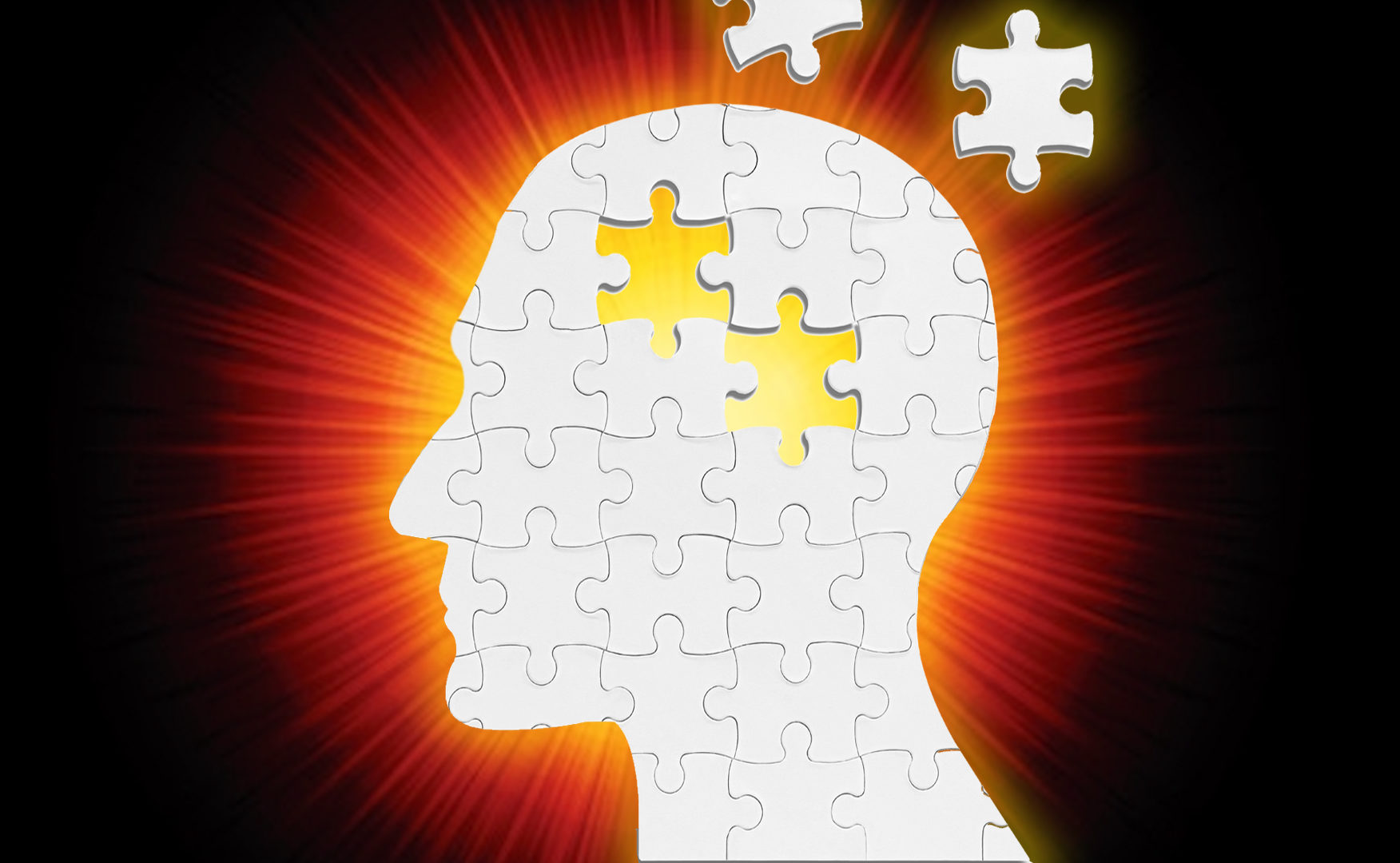

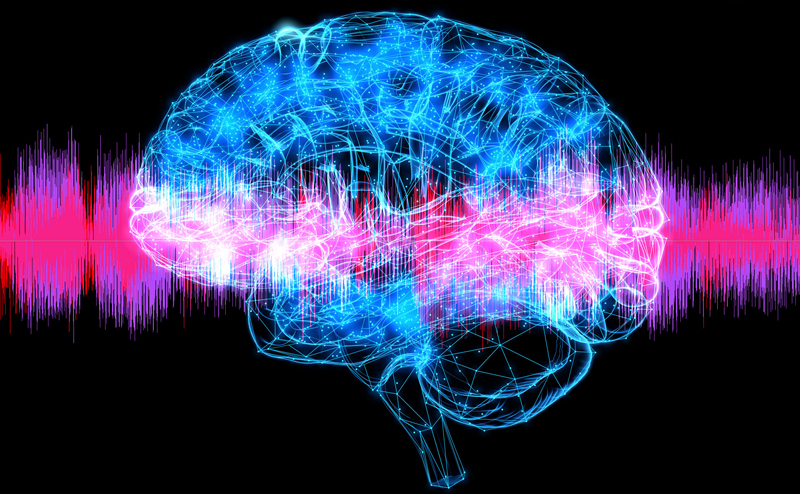
A new generation of technology is revolutionizing neuroscience, allowing a closer study of the brain than had ever seemed possible. The techniques are hybrids of optics, genetics, and synthetic biology with the ability to manipulate brain activity, often in real time. Through direct stimulation of neural connections, some of these techniques hold the promise for the treatment of diseases like depression or schizophrenia.
This program is part of the Big Ideas Series, made possible with support from the John Templeton Foundation.
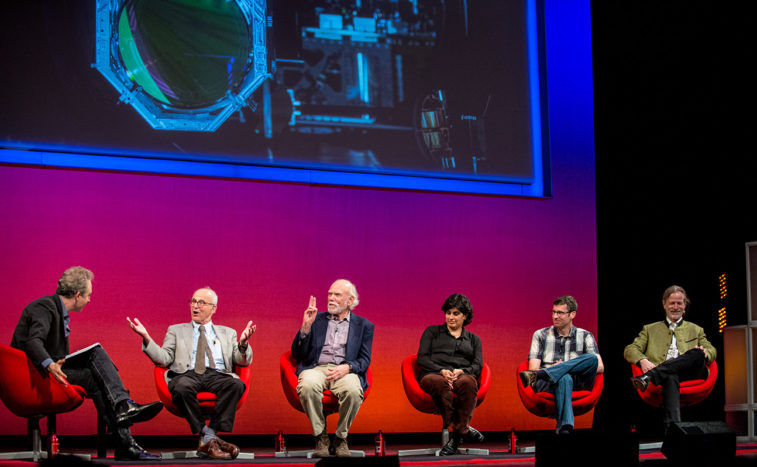
GUY MCKHANN: So I’m Guy McKhann: as I said I’m a neurosurgeon at Columbia. I one of the things I get to do is I specialize in awake brain surgery so I get to operate on patients while we’re looking at their brain while they’re awake while they’re functioning. And the more you do that the more you realize how little we understand and actually know about the brain. So without further ado we’re really privileged to have a tremendous group of participants.
MCKHANN: First we’ve got. Our first guest is professor of biomedical engineering and radiology at Columbia University a member of the Zuckerman mind brain behavior Institute and Kavli Institute for Brain Science.
MCKHANN: Please welcome Elizabeth Hillman.
MCKHANN: Our next participant is an assistant professor of psychiatry, neuroscience and physiology at the NYU Langone Medical Center who investigates the neural circuitry of mating and aggression in mice.
MCKHANN: Please welcome Dayu Lin.
MCKHANN: Also joining us is a neuroscientist and psychiatrist at Weill Cornell Medical College as a psychiatrist specializing in the treatment of complex mood disorders. Please say hello to Conor Liston.
MCKHANN: And our final participant is a neuroscientist,director of neuroscience at Cold Spring Harbor Laboratory who is interested in how brain circuits give rise to behavior. Please welcome Tony Zador.
MCKHANN: So maybe first we could talk about a little bit the history of where did optogenetics come about from. Then you guys could talk about using a little bit.
TONY ZADOR: So interestingly, actually the history of optogenetics can be traced back to an idea that Francis Crick, who’s famous for the double helical structure of DNA first came up with. He proposed that it would be really convenient if we could activate neurons with light. He proposed that something like 30 years ago. And it was people had tried different approaches and were developing different strategies for trying to make it work and it was really the fortuitous recognition that a protein from algae could be expressed in neurons and endow them with the ability to respond immediately to to to light. And that happened I guess about a little over ten years ago and it transformed how we do neuroscience.
When I started in neuroscience everything in terms of trying to understand behavior was correlational – we saw neurons fire and we saw the behavior and we put them together and we of course all recognize that correlation is not causality, but with without the ability to sort of dissect out the circuits that were responsible for the behavior, all we could do is keep trying to find those associations. And so that happened about I guess 2005 and within a couple of years lots of other, lots of labs it happened. It was a paper from Karl Deisseroth and Ed Boyden. And within a couple of years. Dozens and soon hundreds of labs were using it. So that’s really the basic history.
MCKHANN: So Conor you spent time in Deisseroth lab before moving to Cornell so tell us about how you’ve gotten with optogenetics to what you’re doing with it, some of the potential applications as you see it within your world.
CONOR LISTON: Yeah. So as you guys heard I’m a psychiatrist clinically. And one of the things that really excites me about optogenetics is this potential opportunity to understand the neurobiological basis for mental health and mental illness and hopefully develop new treatments. And I think a big limitation in psychiatry is that the drugs we use, I always like to emphasize that the drugs we use they do work and they’ve been a great benefit for a lot of people, but they’re blunt tools, they bathe the brain and they change some aspects of brain function in a way that that helps but they probably have lots of other unintended side effects. And optogenetics affords us this really unique opportunity to really precisely manipulate specific brain circuits, figure out what’s going wrong and try to fix it. And that’s that’s one thing that really excites me.
MCKHANN: So give us give us an example of how you’ve done that, a specific circuit that you’ve targeted and what you’ve been able to find from that.
LISTON: Yeah, so during my time at Stanford, this is like a collaborative work with several other investigators there, we were really interested in understanding how the brain supports reward seeking behavior that’s a thing that’s obviously altered in the brains of many depressed patients. And we were able to precisely manipulate the activity patterns of specific cells in the region of the brain called the prefrontal cortex and begin to understand how, how it how it regulates the processing of reward related cues and motivates animals, and presumably something similar is happening in people’s brains, motivates their their their desire to work to obtain rewards. And interestingly what we found looks a lot like what what goes wrong in the brains of depressed people there’s this abnormal pattern of increased connectivity in this brain circuit and that seems to be driving some of the the reward processing deficits that these that these rats have and then possibly also people
MCKHANN: Dayu you’re using a lot optogenetics so what are you using it for and what types of questions are you able to ask with optogenetics.
DAYU LIN: Yeah. So I was lucky enough actually to be of the first person that will benefit from the optogenetics. I remember it was still in the days when there was a postdoc. And I had to say optogenetics, although it’s a new technique but the concept wise is is not new. In the sense that neuroscientist have tried very hard for centuries, of trying to manipulate the brains in order to see what happened. So but the conventional manipulation is by passing some currents into the brain so the electrical stimulation and that’s where I started when I trying to study the brain before the optogenetic era.
LIN: Electrical stimulation, there is one deficit of that which is that neurons are not just a cell, which a ball, but they actually have a lot of processes coming out. So when we electrically stimulate it we’re not only stimulated to cell body this ball, but also all the processes coming from it. So potentially the cells which is a far away can also be activated because of this electric stimulation. And optogenetics is advantages because it allows you to really target the cells interesting without affecting those cells it just sending processes to it. That’s just the specificity that makes this tool really unique. And now we’re putting optogenetics into those cells and stimulate.
MCKHANN: So why don’t we look at it if we have the aggression video first
LIN: Yeah. So this is a mouse. There was this part of the brain. It’s a very deep very Ventrell. And we turned down this area and you’ll see that he start to attack this glove. And he really you know that’s not what mouse typically do, so they don’t usually attack with gloves but in this particular case when the light activated it he repeatedly sinks his teeth into this glove. In fact when we take it out this glove is in fact deflated. So and it’s not only gloves. In fact that they would attack other conspecifics including a female mouse that a male will typically would not attack at all. So this inanimate object probably shows the extreme of misbehaviors that this control of this area is just a turn on this attacking instinct immediately.
MCKHANN: So there’s some specificity to the aggression.
LIN: It does. It does. In fact, one of the big questions that we are trying to ask in the lab is when we do activate this area, is this just a biting instinct or it’s actually increasing this aggressive motivation. Of the animals and that’s why we’re designing different behavior task in order to dissociate this part. But that’s exactly to ask is it to just make them bite or actually they are become has this urge to aggress.
ZADOR: Can you get them to attack a cat?
LIN: I haven’t tried that, attack a cat or a rat.
MCKHANN: That’s a much more complicated question.
MCKHANN: So all of you guys are talking about a technique where you’re able to select out a very small specific part of the brain to try to isolate and look for behaviors. Now Elizabeth you’ve been focusing on the one hand on using imaging techniques to look at much broader patterns of activity and larger connectivity patterns between the brain and even whole brain activity in various types of animals. Can you fill us in on some of the things you’ve been doing along those lines?
ELIZABETH HILLMAN: We do a lot of technique development in the lab and I was trained as a physicist so I did a lot of optics and so you know these technologies when they come along they don’t just come along, wonderful people conceive of them and piece them together but then using them and figuring out how to do that is really challenging as well and that’s a really important fact to hear so that little mouse had a thing on its head and that was actually holding an optical fiber that was allowing you to put laser light into the actual brain to turn these cells on and off. And so in our work what we’re really interested in you know how can you how can you do that better. I mean it’s not just that you can turn cells on but you can actually turn cells off as well. And the dream is to have many many different colors so that you can be very selectively turning one thing on and turning one thing off. And that really lets you you know figure out what part each one of those things is playing
HILLMAN: I was thinking of the analogy of sort of what is my role in getting my kids out of the house in the morning. All right well how do you tell that you just throw me in there and make me flash. No but if you take me away and then see what happens when I’m not there. You know it’s quite, quite a helpful way of figuring out what it is exactly that I contribute. And so you know it’s not that much actually
But so so so figuring all of this out I mean for me you know we do our own we do our own experiments as well and we’ll talk about that more. But but figuring out you know working working with you guys you know what exactly experiment do you want to do. What behavior do you want to do. Now which areas of the brain do we have to get to and how do we have to make that all work together so that we don’t have to have 10 lasers and all of this stuff happening at the same time as is one of the things that really gets me going. And then and then and then we combine that with imaging because you know what you’ve seen mostly there is is the read out you know you change something in the brain and you look at the way that the person’s actions change or the animal’s actions change. But we can also simultaneously be looking at what effect that has then on the firing of the other neurons.
HILLMAN: And so we do a lot of work trying to capture activity at the level of a few cells at the level of the whole brain. We look at that activity in flies and worms and and little fish. And then we also do it up at the scale of mice and then sort of project that up to the scale of the human brain cells.
MCKHANN: So this would be a good time to look at the video that you have in terms of patterns of activity because it’s very different from exciting a few neurons with one fiber. You’re looking at what the brain is doing spontaneously in its own pattern of pattern of activity.
HILLMAN: so this is work that we’ve been doing. All right to take takes a little bit of explaining. So what you’re seeing here is actually that the entire top surface of a mouse’s brain and that reveals a whole ton of different areas of the brain that we can look at all at the same time. We’re using something called G camp, which is a calcium sensitive fluorescent protein. So what we’re just talking about was optogenetics where you express based on the genetics of the cells you make the cell produce this light sensitive protein. And one of the other major tools that has really revolutionized neuroscience in the last five to 10 years has been these fluorescent proteins like the glowing jellyfish that that you’ve probably seen or the newspapers with the glowing mice right.
HILLMAN: So these are calcium sensitive fluorophores which means that actually when the neurons fire that they actually flash and we can read that out. OK so it’s sort of the opposite.
HILLMAN: So anyway we’ve been observing this in the mice and we’ve found these amazing patterns of spontaneous activity that tick around in the brain. And what I thought would be fun to show you today is that we do a lot of quantitative analysis on this trying to understand the cadence of these patterns and the locations of these patterns and what do they mean and one of the things that we just were able to do. If you go to the next slide. So we. We found that again it’s hard to see with these patterns. So on the top left is just a still of these kind of weird patterns of activity that we really see coursing through the whole brain.
HILLMAN: And we found that they were sort of coming on and off in different regions of the brain. And so I’m going to play a play a movie here where what we’ve done is rather than just showing you the movie of these patterns that seem to kind of roll around in the brain because there’s different regions that are lighting up one after the other. We’ve actually encoded them in color and also as notes on a piano. So what you’re going to hear is kind of what it sounds like in the brain of a mouse as a mouse is just sitting there resting and this is over pretty much the entire surface of the brain so all the regions are responsible for seeing and hearing and motor actions. This is all done by my student Nick here who is a biomedical engineer slash composer. So if you want to just click the. I’m not getting… there you go. So every time a region lights up you hear a note. Give it a second
HILLMAN: So we use this method just as a way to try to just really capture
MCKHANN: Is that fully automated?
HILLMAN: Sorry?
MCKHANN: Is the relationship between the brain activity and the music fully automated or.
HILLMAN: Yes this is actually using the NAM. Non-active matrix factorization algorithm on this stator and then taking the factors and then putting them into musical notes
HILLMAN: And then if you go to the next slide. So then
MCKHANN: Just before you go there, I would argue relative to what Tony Tony was saying before
MCKHANN: At rest, at least based on the music pattern the mice are quite calm.
HILLMAN: Well after I started doing this analysis I remember I had a big grant on this that I submitted right before Thanksgiving and I was sitting there in thanksgiving and I was I just kept feeling this rhythm of my brain you know and thinking you know if I could just sort of calm down watch some television if I just watch you know 10 Things I hate about you one more time I thought maybe I could get myself into this state. You know what I mean. And so this drew us onto to the next part which is we then gave the animal caffeine.
HILLMAN: OK. So go to the next slide and play the movie on the left so, it takes a minute.
ZADOR: So we could we could play this music to a mouse listening to its own brain.
HILLMAN: We talked about that just recently actually if you can use that as a way to actually. So you want to quickly pay the caffeine one It’s not so different. It’s a little faster. Has some gaps. Feels a little different.
HILLMAN: OK and then so we gave it ketamine. So ketamine is a drug Some people use it recreationally and they shouldn’t. And it’s an anesthetic which really makes you basically unconscious makes you high has various effects. Of making you forget and so on. So that’s the one on the right here and you’ll see it has a really profound effect.
HILLMAN: What’s what’s really interesting about this is that this is a very odd modality that we came up with. we actually just take the data using LEDs and camera.. It’s very low key compared to the really high tech stuff that we also do in the lab. But the reason why I’m really excited about this is I think neuroscience for a long time has zoomed really far in on just just the cells you know looking at single cells and how does cells interact with each other. And when we discovered this activity that’s really everywhere. It really changes the way that I’m now trying to think about you know. The brain is just sort of talking to it. It’s like the Internet. Right. It’s not just you and you sitting there it’s you connected to all your Facebook friends and all of those things kind of interacting together and so we’re just dumb.
HILLMAN: We’re having fun with this of course but we’re really using this now to try to understand how that sort of whole brain contributes to behavior. And particularly here we think about state so a state of being anxious a state of being calm. And really what does that look like and then. How would each one of these react differently if you presented it with an aggressive glove.
MCKHANN: So you’re showing these amazing patterns of how the brain connects and what connects to what. You know Tony you’ve taken a totally different approach to try to study major areas and how what actually connects to what and how we unravel that maybe can you can you explain that for us.
ZADOR: Yeah so to sort of put it in perspective. When the when the optogenetics revolution hit, my lab jumped in whole hog we were already training mice, or at that time rats, to perform what we thought were cognitive tasks and we could talk more about that. But we were training them to listen to sounds and make decisions based on the sounds they heard to get rewards. And we were trying to understand the circuits that were involved in making those decisions. And we had identified certain subsets of neurons, using optogenetics later we had gone beyond simply recording the activity of these neurons and actually shown that one set of neurons, turns out that project to the auditory striatum, were actually really important in carrying information about a sound toward driving movement. But, what I realized from that was that you know we can make guesses about the circuits involved and using optogenetics we can actually test whether our guesses are right for the first time like we could test at the circuit level which subset of neurons within a particular area are driving this behavior versus that behavior and what roles they play.
ZADOR: But the problem was that, we just have to make educated guesses. And one of my students was really fantastic and made a really educated guess and guessed right. And chose this this particular pathway. But other students made less successful guesses. And the cost of guessing wrong is a couple years of work. You you do a set of experiments and if you guessed wrong, the answer was nope. That wasn’t the right guess. And you learn very little
MCKHANN: Time for a new career.
ZADOR: Not my career. As I told you I used to tell people I still tell them who enter my lab I quote from Shrek always there’s a there’s a for those of you my son when he was younger watched Shrek over and over again and there’s a scene where Lord Farquhar sends his knights out to find the princess. And he says this mission is dangerous, many of you may die but that’s a risk I’m prepared to take. So, If we guessed wrong there still ones who come up empty handed. So anyway.
MCKHANN: So how did you make it better for them?
ZADOR: So the so the idea was that we needed a way to to screen through the guesses. We needed to know what the circuit was in order to know what was a reasonable guess. Knowing the circuit would not tell us how it actually works but it would rule out all sorts of possibilities. And the problem was that the tools that were available were either not high enough resolution, didn’t have a single neuron resolution or they were incredibly slow. So that what we needed to know was whether there are neurons, let’s say in my case and the auditory cortex that send their axons to this region and that region and whether there are other neurons that send their neurons here and there. And the problem was there was no way of knowing in sort of a high throughput way what the millions and billions of neurons, well millions and a mouse billions in a human where they send their axons. And so the traditional methods for asking those questions were all based on microscopy. The typical high-resolution method is to take one neuron in one animal fill it with with a fluorescent tracer or some other kind of tracer and then track that trace all throughout the brain. The problem is…one neuron per mouse lots of neurons. That’s very slow. If you try to do more than one neuron the problem is that the processes start overlapping and you can’t resolve where the individual processes from these neurons go.
ZADOR: And so the idea that I had eight years ago now, but it’s finally working was to replace the usual way of visualizing the neurons, which is just to look at a fluorescent dye, with
what has turned out to be a fantastic marker which is DNA or in fact RNA. So what we do is we, we cause each neuron to express a unique random sequence of RNA which then gets transported all throughout the cell.
MCKHANN: So but how do you do that why does a neuron want, not want real neural RNA.
ZADOR: Yeah, in real life most neurons do not express random, in general, you don’t have a lot of random labels floating around your body. In fact the only case in our body that we know of where we have random labels is your immune system where the genetic material gets scrambled each time to make antibodies. So we thought about actually trying, and we’re still kind of working on trying to trick neurons into scrambling their DNA, but an easier way to do that is we can make viruses that are, that each express a unique random thirty nucleotide, thirty letter string of DNA and then we just squirt that into the brain and each neuron takes up one viral particle. Sometimes they take up two and we could talk about technically whether it’s not really a problem. And then those, that it gets amplified within the neuron and gets transported out to the axon.
ZADOR: So now we no longer have to carefully trace each little process, we can just use DNA sequencing technology, which has transformed other fields of biology. We can just just piggyback off of these tremendous advances in DNA sequencing technology which have driven the price of sequencing our genome down to below a thousand bucks. We can we can use literally that same technology to figure out where all these neurons project. So we…
MCKHANN: Every neuron has its owns its own individual..
ZADOR: Every neuron has its own individual label, and it actually I should have shown this picture. It’s inspired actually by an idea that scientists at Harvard, Josh Staines and Jeff Lichtman came up with about 10 years ago, called ‘brainbow’ where they cause each neuron to express not one color but a rainbow of colors. And so basically the idea is to replace a rainbow of colors, it’s hard to read out more than a couple of colors so there wasn’t such a big win but it makes beautiful pictures, to replace those with these sequences. And so now what we do is we inject in to one sight in the brain or in fact we can now tile the entire brain with this virus and figure out where all the neurons in the cortex project to.
So in fact I was looking at your pictures and one of them, we were just now starting to analyze this whole brain connectivity map and it turns out that there are sort of communities of neurons that talk preferentially to each other. And in fact the the communities that we find based on these connectivity actually are very reminiscent of the the sort of different chords especially in the ketamine brain where it looks like there’s there’s sort of within their community there’s a lot of conversation and then it passes the message to the next community.
MCKHANN: So it’s possible that you may be seeing with nerve axonal labeling some of the same things
ZADOR: I think actually the substrate for what you’re seeing is precisely…
HILLMAN: We should hang out.
ZADOR: Yeah.
MCKHANN: So how do you say how do you take that to the next or a different level and say Now we know how things all communicate. How do you start studying what’s normal and what’s abnormal?
ZADOR: Right. So so the idea here is that we can now for a couple thousand bucks figure out the whole connectivity of our brain at, there are some issues about resolution. It’s a single neuron resolution but the spatial resolution is limited. We have some ideas and actually we’re working on methods that give you higher resolution. But the real interesting idea is that we can now compare the wiring diagram of a normal mouse to a mouse that has a genetic deficit that has been associated with the human disease. And there are now dozens or hundreds of these mice these so-called animal models of autism and schizophrenia and depression. So we know that that there are genes that cause that are associated with human conditions. We know that when you put those when you disrupt those genes in mice they disrupt behavior in ways that are sometimes similar or sometimes not. But in many cases we think that what’s going on is that disruption of those genes causes some kind of change in the neural circuit.
ZADOR: But once again the traditional approach to figuring out what that change is to take an educated guess. Burn up a couple of grad students and if you guess right then… well but that is how these things go it’s incredibly frustrating. And so the idea is now we can we can actually take one of these mice and say look here is the disruption we’re not sure which of these disruptions are causal in the behavior. But this totally sort of constrains the set of hypotheses that now physiologists like me and the other people on this stage have to consider.
MCKHANN: So you’re taking a lot of guesswork for the grad students out of picking your project?
ZADOR: That’s right. That’s right that’s really my goal is to sacrifice fewer grad students on the altar of bad guesses,
MCKHANN: Which is very admirable. We would all agree
MCKHANN: Connor. Connor you’ve taken a different approach to try to look at populations of neurons by putting little prisms in the brain and combining it with optogenetics. Can you talk about that and what it allows you to do that’s different from traditional optogenetics.
LISTON: Yes so this work is really similar to what Elizabeth was just describing but it’s kind of on a more microscopic scale. So we’re using the same kind of calcium sensors that fluoresce in proportion to their activity. Like you saw on those beautiful videos that Elizabeth showed us a few minutes ago. But we are looking with single cell precision on a much smaller area of the brain. One of the functions that we’re really interested in is working memory. This basic kind of fundamental form of short term memory that we all use all the time like if we decided we wanted to take take a break now and order some pizza. Working memory is like what would enable us to kind of look up our favorite pizza place on your phone and store the memory of the phone number long enough to dial it. But it goes away rapidly
MCKHANN: It’s already gone away with cell phones.
LISTON: Yeah exactly.
LISTON: Teamwork. And it’s disrupted in depression and in schizophrenia and in many other psychiatric conditions in different and interesting ways. So we think it’s kind of a fundamental process we can show that,
MCKHANN: Can we see this Video
LISTON: that video. So so what you’re seeing here are cells in the prefrontal cortex those black, those black kind of bands are blood vessels. But these are individual neurons, brain cells that are expressing this this fluorescent calcium sensor and that Starry Night effect is basically a a readout of how active each one of these cells is over time. And what you what you don’t see is that this mouse is awake and it’s behaving it’s performing a working memory task while we’re getting this readout. And so what we hope to be able to do is test hypotheses about how particular cell types in this image that you’re seeing here how they interact to encode and maintain a trace of the working memory during a delayed period during the time it would take us to dial the phone number in that analogy and how these different cell types interact to imbue working memory with interesting properties. And then once we understand that like like we were just discussing a minute ago we can we can formulate hypotheses, guesses and actually test them using optogenetics to manipulate the activity of specific cell types and and test whether our predictions are actually true or not.
MCKHANN: So everybody here is using different animal models to study these things to try to come up with information that can help us fundamentally understand human behavior. So maybe as a group, whoever wants to can talk a little bit about about how do we best do that. How do we best do that in a way that we are minimizing the use of animals, being compassionate in that you know, but yet understanding what we need to understand. So what are your thoughts on that?
HILLMAN: We recently started using fruit flies and I don’t feel bad about that at all.
MCKHANN: So tell us what can you learn out of a fruit fly?
HILLMAN: The beauty of the fruit fly model is that you can breed them really really quick. And so these genetic techniques now that are really at the core of neuroscience that let us label the cells different colors and change them, introduce genetic diseases, make them express optogenetic options. You can do that in the space of a week. You can just cross two flies and then you have another fly and it’s got rainbows in its brain and it does all this stuff. For me what’s really nice because I when I build microscopes is what you showed what you saw there was about a ten millimeter field of view which is what we need, but we couldn’t see single cells.
But if you if you have a fly’s brain we can fit that whole thing within our microscopes field of view and so we can see every single cell in the entire brain. And so we do these completely crazy things where we get the fly and we actually have it walking on a ball and we’re imaging it’s whole brain while it’s on a ball and then you can actually puff odors and odors of lady flies and then you can see what it does. And you can see where it crawls and then you’re seeing all of this activity in the brain. You know as it’s doing that and so, are flies the same as humans ? Probably not. They they can fly which we can’t. So I don’t know.
HILLMAN: What we’re trying to do on so many different levels is understand building blocks understand basic principles understand you know how does behavior emerge from all of these little cells flashing on and off and so doing it in that system is a very efficient very good nice way of understanding something at that level and then scaling it up so. So there’s these people in neuroscience working at every single level. What we really need to do is all talk to each other and sort of piece together all of those clues. And we also do the maggot’s as well. I mean it’s gross.
MCKHANN: Dayu. Dayu, you showed us that you work on aggression but you also work on maternal instinct so another thing that’s across the animal kingdom but that we all think of as an incredible human thing but actually it’s much across the animal kingdom so maybe you could talk to us about how you study that sort of thing with something like optogenetics.
LIN: You know they are in my views a lot of social behaviors instinctive behaviors that they’re all controlled by those ancient structures that the mechanisms is very conserved. And the maternal and paternal behaviors and in fact parental behaviors in general in fact is also highly sexual dimorphism. I’m all for them and I’m one of the regions we study is controlling the maternal behaviors. So I to show you a video here just to say that we are not only looking at the years which is fighting but also this is like just a heartwarming videos and look at it.
So this was this is a virgin female that date she doesn’t really care about the pups so much and she doesn’t have much experience with the pups. So this is called a sham sham stimulation. In those imitation there were just the endless areas that we put a puppy in the corner and she basically ignored the pups. The pups is a whipping around probably making other boys sound. Tried to get her attention. So. So this part will be sixty seconds which I guess was just trying to convince you that there’s really nothing happened.
LIN: So now pay attention. So now we start to stimulate this part of the brain Right here. And then. Walked right out. And to bring the PAP .Back home. So they are part of the brain regions which are really just controlling those maternal instincts when it turns out they should be instamom mom.
MCKHANN: So supermom do this all day long.
MCKHANN: Yeah. So before we move because a lot of you guys are doing. Not just mouse modeling but also doing human work too. Can you all comment on because, because I don’t know if anyone here is actually working with it in addition to optogenetics out of the Deisseroth lab, another great development has been this technique CLARITY, the ability to take the brain which is in oblique structure and essentially turned it into a window pane. So can you all talk about how you see that as a tool, the strength of it, and can you potentially merge it with some of these other techniques?
ZADOR: Well I think that. So for those of you who haven’t heard there is a technique that allows, so we’d like to see where all the neurons are we’d like to see how they’re all connected, and what limits it is the fact that the neurons are embedded in all sorts of fat and extracellular stuff. And when you do microscopy it’s very difficult to see clearly what’s going on. And so for several decades people have been trying various methods for clearing tissue for making it transparent. And there have been a couple of major advances in the last couple of years including the CLARITY technique from Deisseroth lab and some other techniques that have made it much easier to see a lot of the brain clearly, to be able to image down one to three millimeters in fact as the limits now have less to do with the actual tissue and more to do with microscopy.
I think that really is making it a lot easier to quickly see what the circuitry is how different parts of the brain are connected, We’re using sort of related techniques. In fact we’re using a technique developed by a guy named Ed Boyden at MIT which in some sense goes one step further it allows you to not only clear the tissue but simultaneously to expand it. So some of the structures we’re interested in, it’s called expansion microscopy surprisingly enough. So some of the structures we’re interested in are below the resolution of light and the usual approach to resolving a structure that’s below the resolution of visible light is to use electron microscopy, which is a lot of work.
MCKHANN: A lot of grad students.
ZADOR: It’s more it’s a, it’s a it’s a million dollar machine and it’s it’s it’s a nightmare at least on this scale first for for applying electron microscopy to sort of macroscopic structures. And so what he had, I mean Ed is actually the co-developer of channel and along with Karl Deisseroth, what Ed realized was that instead of getting, making microscopy better so you could see smaller things. It would be just as good if you could make the smaller things bigger. And so he developed a technique that was really that was inspired by and actually uses literally the stuff that they make diapers out of. And you know how you, for those of you who are familiar with diapers you had liquid will say water to a diaper and it expands. And so he imbeds this this this diaper stuff in between the bits of neuron and then binds every little protein to a bit of it and then expands every protein isotropically in all directions. And so you can get expansion factors that are two and four and now even twenty times the original. So you can take a synapse which is below the resolution of light point below point two microns and make it a three micron, five micron structure which we can easily resolve and I think that that technique is really on the verge of transforming a lot of what we can do.
MCKHANN: Can think, you merge that?
ZADOR: So we are we are actively working with lab to merge our approach. So all of this just to be clear is in fixed tissue and tissue from the brain and after it’s out of the skulls. Optogenetics is fantastic for working with a live animal but then to figure out the circuitry of that animal that what you have to do is take it out and then use sort of these methods so we are barcoding neurons in all the animals you know regenerate to perform a task and then we expand the tissue and do the sequencing in situ so we can actually sequence the barcodes while the bar codes are still inside the brain and it gives us the spatial resolution to actually see which barcodes are at a synapse and actually figure out which neurons are connected to each other neurons. So there are some engineering issues, throughputs, throughput issues but conceptually we can expand an entire brain and figure out the wiring diagram of an entire brain at synoptic resolution. And while we’re at it we can also figure out which genes are being expressed. So this is, you know, one of one of the people in Ed’s lab calls it the Rosetta project because basically we can figure out everything about a brain all the way from activity while the animal is performing a task, the behavior. And then the circuitry and gene expression that cause those things so that that’s where we’re hoping to go with them.
MCKHANN: That’s pretty amazing to think about. So but as you say that’s finding out everything about a mouse brain.
ZADOR: Yeah.
MCKHANN: So we’re talking a lot about you know mouse mice have human behaviors and things. You know, Elizabeth you talk a little bit that you have done a very strong proponent of that we’ve already got great human imaging techniques that you’re using. But also in humans in trying to trying to study the differences and similarities and what we can gain more out of our MRI techniques we already have and how you’re going about that and helping translate ideas.
HILLMAN: Sure. So, so I touched on this a little bit before but you know anybody here had an MRI scan? Have you ever volunteered for a functional MRI scan where they showed you how to do tasks and things like this? OK. So I’ve done lots of them, I fall asleep I’m terrible subject. So we, so you go into an MRI scanner it makes this beautiful image of your brain. And what they discovered about 30 years ago now is if you actually took a sequence of images over time that you could see small changes in the signal in different regions of the brain while you were doing things so like if you actually moved your hand you could find a region of the brain that would have some sort of correlated change in the MRI signal while you were doing that task and we call that BOLD, signal the blood oxygen level dependent signal. And it’s what generates those beautiful movies images that you see on the front of the New York Times saying this is the area of the human brain that reacts when you see a picture of a puppy or something.
ZADOR: The puppy area
HILLMAN: And so you know I did my I did my postdoc up in Boston in one of the centers that actually developed this method. And so right now you know this is the best method that we have for looking at the human brain. It’s noninvasive, any one of you can go sit and do one of these studies and pretend to be a mouse, you know tapping your fingers or being shown pictures. And, and it does a pretty good job it points to this area here, this something happened here. Right? The problem is that that signal actually comes about because there was a change in blood flow in that area. So there’s not really a change in the MRI signal when a neuron fires but there’s a change in the magnetic properties of blood when it becomes oxygenated or deoxygenated. And so that little signal change that you see is actually coming from a change in blood flow to the area. And that’s why it gets a little bit confusing because it’s a little slower than the neural activity because it takes a while for blood flow to change after neurons fire.
And so I’ve spent, in addition to the other work I’ve done, I spent a long time studying this the relationship between neural activity in the brain and the blood flow activity that that kind of accompanies it. Partly initially to help with understanding those fMRI images better, functional MRI because when you’re seeing blood. We wanted to understand what that meant in terms of neurons I could talk about this all day but it CT cause us to have to develop a lot of imaging methods to go in and use microscopes to simultaneously look at neurons firing and blood vessels next to them.
HILLMAN: And I got very interested in it from the standpoint of it’s actually quite surprising that every time or region of your brain has some neural activity that there’s actually a big increase in blood flow in that area and that has to happen for a reason and that has to be an important part of brain health. And so now my work has sort of moved towards not just trying to understand the fMRI signal better but also recognizing that there might be situations in which that relationship between neural activity and blood flow gets disrupted. And if every time you’re hungry you try to eat a sandwich but you miss something’s going to happen you’re going to lose weight you’re not going to perform as well. And so, what, all that stuff I showed you with the coffee and the ketamine I mean the really the core of that study in the beginning was looking at how that neural activity actually was coupled to blood flow changes.
And we’ve got ways now we actually can give drugs that disrupt the relationship between neural activity and blood flow and we can then see what effects do they actually have in real time on neural activity, what effects they have on behavior. So we’re training the animals to sit and actually push a lever, this is one of Nick’s other projects right ,there were we’re try and get them to push a lever disrupt this blood flow relationship and see whether they actually become slower at pushing the lever simply because this is just another aspect of the brain that we really don’t understand.
HILLMAN: Stepping back if we can get a better handle on this relationship between blood flow in your neural activity, it gives us a way to really better use functional MRI and it has, this method has kind of an image problem, right? We all agree. Because it’s not so clear what it means. And in fact when you do it in someone who just had a stroke you might see huge activity in an area where they’re paralyzed or you might see no activity at all and they go on to be, to have a great recovery. And so sort of my mission is to, to use these methods more in the mice where we can do this very detailed imaging but to try to disambiguate all of this. Try to find is there an early signature of Alzheimer’s. Is there something that happens in stroke that we can actually understand in terms of what it’s going to look like in MRI and then can we use fMRI in a much more guided way to get to those conditions in human brain. So sounds a little circuitous but it keeps me busy.
MCKHANN: I think we all agree it’s something there’s a huge need for, to understand what is it we’re seeing and how do we learn from it. You know you’ve done some of the same thing in trying to tease apart subtypes of depression using, using functional imaging. Can you tell us kind of how you approach that and what you’ve found and what you think the potential for that kind of analysis is.
LISTON: Yeah, yeah. We’re really excited about, this is another area we’re really excited about in my lab the opportunity to potentially use brain imaging to rethink the way we diagnose psychiatry, if you make diagnoses in psychiatry. If you’ll bear with me for like two minutes I’ll tell you an interesting historical like little background story. So our DSM, this book that we use for diagnosis is kind of seeped into the public lexicon right, you read about in the Times a lot of people are familiar with it. And the whole way we diagnose mental illness is really not very old, it was completely changed in 1980. I was born in 1980. You can’t say that about very many other areas of medicine. And yet it’s the truth. Everything we do in psychiatry is quite different from before 1980, due in part to this kind of controversial study that was published in Science seven years before that just showed essentially that are our diagnostic system at that time was not very reproducible and very prone to bias. So social psychologist at Stanford very cleverly designed a set of experiments where he sent his investigators into hospitals around the country presenting with a fake symptom. They said I’m experiencing voices saying the words ‘empty’, ‘hollow’ or ‘thud’ and otherwise they were supposed to answer all questions honestly truthfully and and the results they just followed them and the results were really shocking to everybody. They got different diagnoses in different places and later work showed that your socioeconomic background and your ethnic background were could bias the way people assigned diagnoses to you. And, and it was really hard to detect that these were fake patients basically that’s, that’s what he showed you could bias people’s judgment in that way.
LISTON: And then in the second part of this study, and this is kind of the clincher, in the second part of the study the lead of this study had met the director of a famous hospital at the time and he said you know this is this is an anomaly what you found is not representative of the way we do business. Come to my hospital see if you can see if you’ll get different results. And so they agreed he would send an unknown number of people into his hospital over the next few months and during that time their job was to figure out who is a fake patient who is a real patient. Twenty percent of people were identified as definite impostures and other twenty percent were identified as probable impostures. So fully two out of every five people who came to the hospital had, were thought to be possible impostures. And the truth was that he hadn’t sent any impostors and that they were all real patients. So you could bias people’s judgments in both ways.
And so getting to that point here, the DSM3 which was released in 1980 and very similar to the one we use today was really designed to lump lots of people together with very different problems in big diagnostic categories that we could all agree on. And it did a really good job of that. The diagnoses are much more reproducible than they used to be, less prone to bias. But the problem is they don’t correspond to biology very well.
LISTON: So, depression is a great example of that. You have this like choose five or more of nine symptoms. So there’s at least two hundred fifty six ways you can be depressed. And some of them are almost opposites of one another like sleeping nineteen hours a day, can’t get out of bed. Not enjoying any activities, very slowed, gained a lot of weight. That person is depressed. So is a person who’s sleeping three hours a day anxious, weight loss, almost the opposite. And they get the same diagnoses but they probably don’t have the same biological problem and it’s a miracle really that treatments work as well as they do in these in these very different people. So what we’re trying to do with fMRI is figure out whether we can identify clusters of patients that have similar biological problems as indexed by fMRI and which can be used to map functional connections in the brain. And if so then maybe we can then maybe we can target specific treatments to individual people who are most likely to benefit from that.
MCKHANN: And can you find different patterns of patients based on fMRI?
LISTON: Yeah. So we’ve found, I always emphasize I don’t think this is the best or final solution to this problem but it’s it’s a it’s one solution that works well we’ve found, that we can identify four very different patterns of abnormal functional connectivity in the brains of different depressed people. We can assign individual people to those categories based just on their brain scan and then those, those categories predict different kinds of symptoms and a different likelihood of responding to transcranial magnetic stimulation which is a brain stimulation based treatment that you could imagine, I mean it it’s on a much coarser scale, but the goal is the same as with optogenetics where you’re trying to stimulate a particular circuit to to modify the activity of that circuits. TCS works in much the same way and it’s and it’s much more effective in one of these subtypes than in than in the others.
MCKHANN: And do you think if you look for those patterns in mice you could find out for Tony if mice are depressed.
LISTON: Yeah where we’re working on that. One thing I think that’s really nice about the mice is that you can, you know, Elizabeth talked about this. The the one of the limits of MRI is it’s fundamentally kind of correlational in nature. You observe this change in the brain scan and it seems to correlate with a particular behavior or a symptom. But there are many changes in these brain scans and it’s really hard to pinpoint which one if any is causally related to particular behaviors and mice afford us that that opportunity. So that’s that’s one thing we’re trying to do in the brains of our, of our mice.
MCKHANN: So, we want to leave a little bit of time for questions, but so, one final thought for anyone here, so looking down the road, five years down the road what’s your wish list or what’s your vision of where you think we’re going to be or where do you think is going to be the next? What are we going to do to unlock this and take it the next step and what are what are we going to be talking about a decade from now?
ZADOR: Well I can tell you what we’re trying to do. I laid it out. It’s the Rosetta brain. I think being able to understand behavior all the way through from the behavior to the neural activity to the actual wiring diagram for me is sort of the Holy Grail. To to reduce, it’s not even to reduce, it’s to provide the foundation. I went into neuroscience because I wanted to understand how three pounds of stuff gives everything that we see, hear and feel and it just seemed incredibly mysterious to me. And after being in a neuroscientist for several decades it remains just as mysterious. I don’t think we’re any closer to…
MCKHANN: A little bit?
ZADOR: No sadly.
ZADOR: But I mean the core question, why does it how does it…. why are we conscious? How do we even how does it feel. How is it that we feel the way we do? I don’t think we’re any closer to understanding. And I sat there thinking about it for a long time and decided that thinking about it wasn’t going to get us anywhere further. But I’m hoping that if we actually see an example where we can go all the way from the neural activity to the, through the behavior to the circuitry, it’ll at least provide us a way of maybe formulating the right next question which I currently don’t have.
ZADOR: My Ph.D. adviser was Christof Koch who has spent most of his career trying to understand consciousness. And I think now he he’s the scientific director at the Allen Institute where their goals are very similar to the goals of the people here which is to tease apart the circuitry of a mouse. And so my my feeling is that although the, the circuitry of a mouse is clearly different from the circuitry of a human, basically the building blocks are already there by the time you get to the cortex. If you look at a piece of mouse cortex and you change the scale bar and you put it next to a piece of human cortex, they are indistinguishable.
So the basic building blocks we think, that you’re smiling because you’re a neurosurgeon or you will not be able to tell the difference if I change the scale bar – even the neuroanatomists at the Allen Institute were fooled by this this pair of pictures. So I think that ,you know, we as as humans evolved in the last couple of hundred thousand, maybe million years as primates over the last couple of million. I mean most of the work was done I think by the time we evolved as mammals. We split off from mice seventy, eighty million years ago. The basic building blocks I think are there for us to understand. And once we get those, than I think we’re off to the races to figure out what makes us human. But I would be, I would be pretty happy if we could get an understanding of what makes us mammal.
MCKHANN: So we want to learn a little bit of time of questions for questions We’ve got two microphones going around so if you have a question please wait till somebody brings you a microphone.
AUDIENCE #1: Thank you very much to the panel. You’ve given a really great overview of some of the genetic and biochemical techniques that have advanced the field in the last decade or so. But can you talk more to some of the informatic techniques that might be advancing the field you mentioned NMF for dimensionality reduction. There have been issues with, you know far more specifically with sort of the kernels and statistics that are going on there. But how has you know Big Data, data science, machine learning also influenced the field Thank you.
ZADOR: Well I’ll just say that I started off in machine learning neural networks as a grad student and I think that those that field has affected it in two ways. First of all it inspires us how to think about how the brain works. Trying to build something is one of the best ways of figuring out how something works. Beyond that, in the last couple of years machine learning techniques have transformed big data in neuroscience just like they’ve, they’re transforming it in all other, in many other fields. So that our ability, I mean we’re now collecting huge data sets and, you know, either calcium imaging or connectivity, and, you know, you can’t you can’t just stare at it anymore it’s the same question of, machine learning approaches provide ways of generating hypotheses again, just just like knowing the connectivity. And then it takes a real brain at least so far, we’re not quite out of business yet, but these these big data techniques transform, have are in the process of transforming how we sort of generate scientific hypotheses that then we can go out and test. At least for me that’s one of the big ways
HILLMAN: I would say coming back again to the other point is, we need computer scientists and people who understand code and big data to understand the brain. Right. Because it is a computer right? And again the insights you get from people who who who know that are really critical. What’s really fascinating we’ve got Amazon and Google and all of those big companies now really interested in brain research. They’re hiring our graduate students, right? This deep learning, these algorithms. I mean the whole thing is coming together. So so I think both fields are now really stimulating each other and I think you know that’s going to be a path towards getting where we need to go.
LIN: Part of the difficulty is to understand, actually the behaviors. And the behaviors, it turned that a machine can do a better job than humans in order to determine how many forms of a behaviors they are. Actually Bob Datta in Harvard, they are creating a syllable of the behaviors and they’re trying to find a neural correlate to that, which turned out, you actually finding exact in neural correlate to the syllables of the behaviors, which we as a human observer you could have completely missing it. So I think that’s kind of how these two things are neuroscience and machine learning aspects are really assisting each other.
MCKHANN: Another question?
AUDIENCE #2: Depressed lab animals produce, developed isolated, do you alter the genetics?
MCKHANN: How do you produce a depressed animal?
LISTON: Yeah I can take that one. The real challenge you mentioned like th, like animal models of mouse models of schizophrenia, autism, we have a much better handle on the genetics of some psychiatric disorders like schizophrenia and autism than we do for depression. What we know about depression is it’s it’s it’s heritable. If your mom is depressed you’re much more likely to grow up to be depressed yourself. And that’s not just about environment. But it’s probably the result of the shared impact of many many genes each having a very small influence on your likelihood of becoming depression and that interacting with your experience. So that makes it a lot harder to have like a genetically-based model of depression that they just don’t exist yet. They may one day
ZADOR: I’ll push back on that a little. There’s a, there’s a model of learned…one model of depression as a model of cold learned helplessness. And there was a guy who bred generation after generation rats that showed this this trait of learned helplessness and over the last, I think it took him a good fifteen, twenty years to breed these. Unfortunately he started doing this when rats seemed like they were going to be the right model system not mice. But he bred these rats that were genetically distinct and showed this trait of learned helplessness and they, he and others have gone on to dissect some of the circuitry that is disrupted in these and identified the lateral habenula, the particularly important circuit in this. Now that’s one particular type of depression. But I think it just shows how the kinds of behaviors that we have can actually often be modeled in a rodent..
ZADOR: And I’ll just also add to that to say that you can even study things like confidence in a rat or a mouse. It’s not obvious that something like confidence, which feels like it’s an internal state would have a neural correlate. But one of my colleagues at Cold Spring Harbor is identified a brain region and particular types of neurons that seem to encode an animal’s confidence and confidence is for example one of the things that can be easily disrupted in depression. So I think that when you actually really think hard about how to probe that behavior in a way that, traditionally, people who work on rats and mice have not but are now starting to, you can actually sort of get more traction on these things than you might have thought. The sort of ridiculous notion of you know a depressed mouse or rat becomes less ridiculous.
MCKHANN: Another question.
AUDIENCE #3: Thank you for the presentation. You talked a little bit about the advance how these advances impact the diagnosis of mental disorders. How do you see on the practical level, what do you see these advances such as optogenetics or anything else that you talked about here, how do you see them advancing the treatment? Like what can we expect in the next couple of years with things like ECT or anything that I might not even be able to understand from your answer to this. Thank you.
LISTON: I think getting back to the transcranial magnetic stimulation thing as one example of a treatment that’s based on stimulating a brain region is, electroconvulsive therapy is is is our most effective by far treatment for depression. And it’s basically a big jolt that is to the brain that’s very stimulating to everywhere in the brain. Right? And and yet it also has a lot of undesirable side effects. And so we reserve it for people who are most who most need it, who don’t respond well to other treatments. I think one potentially exciting avenue in future would be figuring out whether there are particular more focal brain areas that require a particular kind of stimulation. You could even imagine maybe a little further down the road, cell type specific stimulation getting with a lot of Tony’s work was describing. The overall goal being more precision in the way where we’re stimulating brain regions might lead to more effective antidepressant responses and fewer side effects. And I think that’s a vision that everyone kind of shares.
MCKHANN:So unfortunately we are running out of time. We really need to thank these amazing scientists for being here.
A new generation of technology is revolutionizing neuroscience, allowing a closer study of the brain than had ever seemed possible. The techniques are hybrids of optics, genetics, and synthetic biology with the ability to manipulate brain activity, often in real time. Through direct stimulation of neural connections, some of these techniques hold the promise for the treatment of diseases like depression or schizophrenia.
This program is part of the Big Ideas Series, made possible with support from the John Templeton Foundation.

Guy McKhann has over 20 years of experience as a neurosurgeon/neuroscientist at Columbia University/New York Presbyterian Hospital, combining clinical skill and compassionate care to maximize patient outcomes.
Read More
In 2001, Dayu Lin received a B.S. in biological sciences from Fudan University in China. In 2006, she received her Ph.D. in Neurobiology from Duke University, working with Larry Katz. She studied as a postdoctoral fellow with David Anderson at California Institute of Technology.
Read More
Dr. Elizabeth Hillman is a Professor of Biomedical Engineering and Radiology at Columbia University, and a member of the Zuckerman Mind Brain Behavior Institute and Kavli Institute for Brain Science.
Read More
Conor Liston is a neuroscientist and psychiatrist at Weill Cornell Medical College, and his laboratory operates at the interface between these two fields. He leads a team of scientists who are working to advance our understanding of how brain circuits support learning, memory, and motivation, and how these processes are disrupted in depression, autism, and other neuropsychiatric illnesses.
Read More
Anthony Zador is a neuroscientist interested in how brain circuits give rise to behavior, and how disruption of brain circuits can lead to neuropsychiatric disorders like autism, depression, and schizophrenia.
Read MoreGUY MCKHANN: So I’m Guy McKhann: as I said I’m a neurosurgeon at Columbia. I one of the things I get to do is I specialize in awake brain surgery so I get to operate on patients while we’re looking at their brain while they’re awake while they’re functioning. And the more you do that the more you realize how little we understand and actually know about the brain. So without further ado we’re really privileged to have a tremendous group of participants.
MCKHANN: First we’ve got. Our first guest is professor of biomedical engineering and radiology at Columbia University a member of the Zuckerman mind brain behavior Institute and Kavli Institute for Brain Science.
MCKHANN: Please welcome Elizabeth Hillman.
MCKHANN: Our next participant is an assistant professor of psychiatry, neuroscience and physiology at the NYU Langone Medical Center who investigates the neural circuitry of mating and aggression in mice.
MCKHANN: Please welcome Dayu Lin.
MCKHANN: Also joining us is a neuroscientist and psychiatrist at Weill Cornell Medical College as a psychiatrist specializing in the treatment of complex mood disorders. Please say hello to Conor Liston.
MCKHANN: And our final participant is a neuroscientist,director of neuroscience at Cold Spring Harbor Laboratory who is interested in how brain circuits give rise to behavior. Please welcome Tony Zador.
MCKHANN: So maybe first we could talk about a little bit the history of where did optogenetics come about from. Then you guys could talk about using a little bit.
TONY ZADOR: So interestingly, actually the history of optogenetics can be traced back to an idea that Francis Crick, who’s famous for the double helical structure of DNA first came up with. He proposed that it would be really convenient if we could activate neurons with light. He proposed that something like 30 years ago. And it was people had tried different approaches and were developing different strategies for trying to make it work and it was really the fortuitous recognition that a protein from algae could be expressed in neurons and endow them with the ability to respond immediately to to to light. And that happened I guess about a little over ten years ago and it transformed how we do neuroscience.
When I started in neuroscience everything in terms of trying to understand behavior was correlational – we saw neurons fire and we saw the behavior and we put them together and we of course all recognize that correlation is not causality, but with without the ability to sort of dissect out the circuits that were responsible for the behavior, all we could do is keep trying to find those associations. And so that happened about I guess 2005 and within a couple of years lots of other, lots of labs it happened. It was a paper from Karl Deisseroth and Ed Boyden. And within a couple of years. Dozens and soon hundreds of labs were using it. So that’s really the basic history.
MCKHANN: So Conor you spent time in Deisseroth lab before moving to Cornell so tell us about how you’ve gotten with optogenetics to what you’re doing with it, some of the potential applications as you see it within your world.
CONOR LISTON: Yeah. So as you guys heard I’m a psychiatrist clinically. And one of the things that really excites me about optogenetics is this potential opportunity to understand the neurobiological basis for mental health and mental illness and hopefully develop new treatments. And I think a big limitation in psychiatry is that the drugs we use, I always like to emphasize that the drugs we use they do work and they’ve been a great benefit for a lot of people, but they’re blunt tools, they bathe the brain and they change some aspects of brain function in a way that that helps but they probably have lots of other unintended side effects. And optogenetics affords us this really unique opportunity to really precisely manipulate specific brain circuits, figure out what’s going wrong and try to fix it. And that’s that’s one thing that really excites me.
MCKHANN: So give us give us an example of how you’ve done that, a specific circuit that you’ve targeted and what you’ve been able to find from that.
LISTON: Yeah, so during my time at Stanford, this is like a collaborative work with several other investigators there, we were really interested in understanding how the brain supports reward seeking behavior that’s a thing that’s obviously altered in the brains of many depressed patients. And we were able to precisely manipulate the activity patterns of specific cells in the region of the brain called the prefrontal cortex and begin to understand how, how it how it regulates the processing of reward related cues and motivates animals, and presumably something similar is happening in people’s brains, motivates their their their desire to work to obtain rewards. And interestingly what we found looks a lot like what what goes wrong in the brains of depressed people there’s this abnormal pattern of increased connectivity in this brain circuit and that seems to be driving some of the the reward processing deficits that these that these rats have and then possibly also people
MCKHANN: Dayu you’re using a lot optogenetics so what are you using it for and what types of questions are you able to ask with optogenetics.
DAYU LIN: Yeah. So I was lucky enough actually to be of the first person that will benefit from the optogenetics. I remember it was still in the days when there was a postdoc. And I had to say optogenetics, although it’s a new technique but the concept wise is is not new. In the sense that neuroscientist have tried very hard for centuries, of trying to manipulate the brains in order to see what happened. So but the conventional manipulation is by passing some currents into the brain so the electrical stimulation and that’s where I started when I trying to study the brain before the optogenetic era.
LIN: Electrical stimulation, there is one deficit of that which is that neurons are not just a cell, which a ball, but they actually have a lot of processes coming out. So when we electrically stimulate it we’re not only stimulated to cell body this ball, but also all the processes coming from it. So potentially the cells which is a far away can also be activated because of this electric stimulation. And optogenetics is advantages because it allows you to really target the cells interesting without affecting those cells it just sending processes to it. That’s just the specificity that makes this tool really unique. And now we’re putting optogenetics into those cells and stimulate.
MCKHANN: So why don’t we look at it if we have the aggression video first
LIN: Yeah. So this is a mouse. There was this part of the brain. It’s a very deep very Ventrell. And we turned down this area and you’ll see that he start to attack this glove. And he really you know that’s not what mouse typically do, so they don’t usually attack with gloves but in this particular case when the light activated it he repeatedly sinks his teeth into this glove. In fact when we take it out this glove is in fact deflated. So and it’s not only gloves. In fact that they would attack other conspecifics including a female mouse that a male will typically would not attack at all. So this inanimate object probably shows the extreme of misbehaviors that this control of this area is just a turn on this attacking instinct immediately.
MCKHANN: So there’s some specificity to the aggression.
LIN: It does. It does. In fact, one of the big questions that we are trying to ask in the lab is when we do activate this area, is this just a biting instinct or it’s actually increasing this aggressive motivation. Of the animals and that’s why we’re designing different behavior task in order to dissociate this part. But that’s exactly to ask is it to just make them bite or actually they are become has this urge to aggress.
ZADOR: Can you get them to attack a cat?
LIN: I haven’t tried that, attack a cat or a rat.
MCKHANN: That’s a much more complicated question.
MCKHANN: So all of you guys are talking about a technique where you’re able to select out a very small specific part of the brain to try to isolate and look for behaviors. Now Elizabeth you’ve been focusing on the one hand on using imaging techniques to look at much broader patterns of activity and larger connectivity patterns between the brain and even whole brain activity in various types of animals. Can you fill us in on some of the things you’ve been doing along those lines?
ELIZABETH HILLMAN: We do a lot of technique development in the lab and I was trained as a physicist so I did a lot of optics and so you know these technologies when they come along they don’t just come along, wonderful people conceive of them and piece them together but then using them and figuring out how to do that is really challenging as well and that’s a really important fact to hear so that little mouse had a thing on its head and that was actually holding an optical fiber that was allowing you to put laser light into the actual brain to turn these cells on and off. And so in our work what we’re really interested in you know how can you how can you do that better. I mean it’s not just that you can turn cells on but you can actually turn cells off as well. And the dream is to have many many different colors so that you can be very selectively turning one thing on and turning one thing off. And that really lets you you know figure out what part each one of those things is playing
HILLMAN: I was thinking of the analogy of sort of what is my role in getting my kids out of the house in the morning. All right well how do you tell that you just throw me in there and make me flash. No but if you take me away and then see what happens when I’m not there. You know it’s quite, quite a helpful way of figuring out what it is exactly that I contribute. And so you know it’s not that much actually
But so so so figuring all of this out I mean for me you know we do our own we do our own experiments as well and we’ll talk about that more. But but figuring out you know working working with you guys you know what exactly experiment do you want to do. What behavior do you want to do. Now which areas of the brain do we have to get to and how do we have to make that all work together so that we don’t have to have 10 lasers and all of this stuff happening at the same time as is one of the things that really gets me going. And then and then and then we combine that with imaging because you know what you’ve seen mostly there is is the read out you know you change something in the brain and you look at the way that the person’s actions change or the animal’s actions change. But we can also simultaneously be looking at what effect that has then on the firing of the other neurons.
HILLMAN: And so we do a lot of work trying to capture activity at the level of a few cells at the level of the whole brain. We look at that activity in flies and worms and and little fish. And then we also do it up at the scale of mice and then sort of project that up to the scale of the human brain cells.
MCKHANN: So this would be a good time to look at the video that you have in terms of patterns of activity because it’s very different from exciting a few neurons with one fiber. You’re looking at what the brain is doing spontaneously in its own pattern of pattern of activity.
HILLMAN: so this is work that we’ve been doing. All right to take takes a little bit of explaining. So what you’re seeing here is actually that the entire top surface of a mouse’s brain and that reveals a whole ton of different areas of the brain that we can look at all at the same time. We’re using something called G camp, which is a calcium sensitive fluorescent protein. So what we’re just talking about was optogenetics where you express based on the genetics of the cells you make the cell produce this light sensitive protein. And one of the other major tools that has really revolutionized neuroscience in the last five to 10 years has been these fluorescent proteins like the glowing jellyfish that that you’ve probably seen or the newspapers with the glowing mice right.
HILLMAN: So these are calcium sensitive fluorophores which means that actually when the neurons fire that they actually flash and we can read that out. OK so it’s sort of the opposite.
HILLMAN: So anyway we’ve been observing this in the mice and we’ve found these amazing patterns of spontaneous activity that tick around in the brain. And what I thought would be fun to show you today is that we do a lot of quantitative analysis on this trying to understand the cadence of these patterns and the locations of these patterns and what do they mean and one of the things that we just were able to do. If you go to the next slide. So we. We found that again it’s hard to see with these patterns. So on the top left is just a still of these kind of weird patterns of activity that we really see coursing through the whole brain.
HILLMAN: And we found that they were sort of coming on and off in different regions of the brain. And so I’m going to play a play a movie here where what we’ve done is rather than just showing you the movie of these patterns that seem to kind of roll around in the brain because there’s different regions that are lighting up one after the other. We’ve actually encoded them in color and also as notes on a piano. So what you’re going to hear is kind of what it sounds like in the brain of a mouse as a mouse is just sitting there resting and this is over pretty much the entire surface of the brain so all the regions are responsible for seeing and hearing and motor actions. This is all done by my student Nick here who is a biomedical engineer slash composer. So if you want to just click the. I’m not getting… there you go. So every time a region lights up you hear a note. Give it a second
HILLMAN: So we use this method just as a way to try to just really capture
MCKHANN: Is that fully automated?
HILLMAN: Sorry?
MCKHANN: Is the relationship between the brain activity and the music fully automated or.
HILLMAN: Yes this is actually using the NAM. Non-active matrix factorization algorithm on this stator and then taking the factors and then putting them into musical notes
HILLMAN: And then if you go to the next slide. So then
MCKHANN: Just before you go there, I would argue relative to what Tony Tony was saying before
MCKHANN: At rest, at least based on the music pattern the mice are quite calm.
HILLMAN: Well after I started doing this analysis I remember I had a big grant on this that I submitted right before Thanksgiving and I was sitting there in thanksgiving and I was I just kept feeling this rhythm of my brain you know and thinking you know if I could just sort of calm down watch some television if I just watch you know 10 Things I hate about you one more time I thought maybe I could get myself into this state. You know what I mean. And so this drew us onto to the next part which is we then gave the animal caffeine.
HILLMAN: OK. So go to the next slide and play the movie on the left so, it takes a minute.
ZADOR: So we could we could play this music to a mouse listening to its own brain.
HILLMAN: We talked about that just recently actually if you can use that as a way to actually. So you want to quickly pay the caffeine one It’s not so different. It’s a little faster. Has some gaps. Feels a little different.
HILLMAN: OK and then so we gave it ketamine. So ketamine is a drug Some people use it recreationally and they shouldn’t. And it’s an anesthetic which really makes you basically unconscious makes you high has various effects. Of making you forget and so on. So that’s the one on the right here and you’ll see it has a really profound effect.
HILLMAN: What’s what’s really interesting about this is that this is a very odd modality that we came up with. we actually just take the data using LEDs and camera.. It’s very low key compared to the really high tech stuff that we also do in the lab. But the reason why I’m really excited about this is I think neuroscience for a long time has zoomed really far in on just just the cells you know looking at single cells and how does cells interact with each other. And when we discovered this activity that’s really everywhere. It really changes the way that I’m now trying to think about you know. The brain is just sort of talking to it. It’s like the Internet. Right. It’s not just you and you sitting there it’s you connected to all your Facebook friends and all of those things kind of interacting together and so we’re just dumb.
HILLMAN: We’re having fun with this of course but we’re really using this now to try to understand how that sort of whole brain contributes to behavior. And particularly here we think about state so a state of being anxious a state of being calm. And really what does that look like and then. How would each one of these react differently if you presented it with an aggressive glove.
MCKHANN: So you’re showing these amazing patterns of how the brain connects and what connects to what. You know Tony you’ve taken a totally different approach to try to study major areas and how what actually connects to what and how we unravel that maybe can you can you explain that for us.
ZADOR: Yeah so to sort of put it in perspective. When the when the optogenetics revolution hit, my lab jumped in whole hog we were already training mice, or at that time rats, to perform what we thought were cognitive tasks and we could talk more about that. But we were training them to listen to sounds and make decisions based on the sounds they heard to get rewards. And we were trying to understand the circuits that were involved in making those decisions. And we had identified certain subsets of neurons, using optogenetics later we had gone beyond simply recording the activity of these neurons and actually shown that one set of neurons, turns out that project to the auditory striatum, were actually really important in carrying information about a sound toward driving movement. But, what I realized from that was that you know we can make guesses about the circuits involved and using optogenetics we can actually test whether our guesses are right for the first time like we could test at the circuit level which subset of neurons within a particular area are driving this behavior versus that behavior and what roles they play.
ZADOR: But the problem was that, we just have to make educated guesses. And one of my students was really fantastic and made a really educated guess and guessed right. And chose this this particular pathway. But other students made less successful guesses. And the cost of guessing wrong is a couple years of work. You you do a set of experiments and if you guessed wrong, the answer was nope. That wasn’t the right guess. And you learn very little
MCKHANN: Time for a new career.
ZADOR: Not my career. As I told you I used to tell people I still tell them who enter my lab I quote from Shrek always there’s a there’s a for those of you my son when he was younger watched Shrek over and over again and there’s a scene where Lord Farquhar sends his knights out to find the princess. And he says this mission is dangerous, many of you may die but that’s a risk I’m prepared to take. So, If we guessed wrong there still ones who come up empty handed. So anyway.
MCKHANN: So how did you make it better for them?
ZADOR: So the so the idea was that we needed a way to to screen through the guesses. We needed to know what the circuit was in order to know what was a reasonable guess. Knowing the circuit would not tell us how it actually works but it would rule out all sorts of possibilities. And the problem was that the tools that were available were either not high enough resolution, didn’t have a single neuron resolution or they were incredibly slow. So that what we needed to know was whether there are neurons, let’s say in my case and the auditory cortex that send their axons to this region and that region and whether there are other neurons that send their neurons here and there. And the problem was there was no way of knowing in sort of a high throughput way what the millions and billions of neurons, well millions and a mouse billions in a human where they send their axons. And so the traditional methods for asking those questions were all based on microscopy. The typical high-resolution method is to take one neuron in one animal fill it with with a fluorescent tracer or some other kind of tracer and then track that trace all throughout the brain. The problem is…one neuron per mouse lots of neurons. That’s very slow. If you try to do more than one neuron the problem is that the processes start overlapping and you can’t resolve where the individual processes from these neurons go.
ZADOR: And so the idea that I had eight years ago now, but it’s finally working was to replace the usual way of visualizing the neurons, which is just to look at a fluorescent dye, with
what has turned out to be a fantastic marker which is DNA or in fact RNA. So what we do is we, we cause each neuron to express a unique random sequence of RNA which then gets transported all throughout the cell.
MCKHANN: So but how do you do that why does a neuron want, not want real neural RNA.
ZADOR: Yeah, in real life most neurons do not express random, in general, you don’t have a lot of random labels floating around your body. In fact the only case in our body that we know of where we have random labels is your immune system where the genetic material gets scrambled each time to make antibodies. So we thought about actually trying, and we’re still kind of working on trying to trick neurons into scrambling their DNA, but an easier way to do that is we can make viruses that are, that each express a unique random thirty nucleotide, thirty letter string of DNA and then we just squirt that into the brain and each neuron takes up one viral particle. Sometimes they take up two and we could talk about technically whether it’s not really a problem. And then those, that it gets amplified within the neuron and gets transported out to the axon.
ZADOR: So now we no longer have to carefully trace each little process, we can just use DNA sequencing technology, which has transformed other fields of biology. We can just just piggyback off of these tremendous advances in DNA sequencing technology which have driven the price of sequencing our genome down to below a thousand bucks. We can we can use literally that same technology to figure out where all these neurons project. So we…
MCKHANN: Every neuron has its owns its own individual..
ZADOR: Every neuron has its own individual label, and it actually I should have shown this picture. It’s inspired actually by an idea that scientists at Harvard, Josh Staines and Jeff Lichtman came up with about 10 years ago, called ‘brainbow’ where they cause each neuron to express not one color but a rainbow of colors. And so basically the idea is to replace a rainbow of colors, it’s hard to read out more than a couple of colors so there wasn’t such a big win but it makes beautiful pictures, to replace those with these sequences. And so now what we do is we inject in to one sight in the brain or in fact we can now tile the entire brain with this virus and figure out where all the neurons in the cortex project to.
So in fact I was looking at your pictures and one of them, we were just now starting to analyze this whole brain connectivity map and it turns out that there are sort of communities of neurons that talk preferentially to each other. And in fact the the communities that we find based on these connectivity actually are very reminiscent of the the sort of different chords especially in the ketamine brain where it looks like there’s there’s sort of within their community there’s a lot of conversation and then it passes the message to the next community.
MCKHANN: So it’s possible that you may be seeing with nerve axonal labeling some of the same things
ZADOR: I think actually the substrate for what you’re seeing is precisely…
HILLMAN: We should hang out.
ZADOR: Yeah.
MCKHANN: So how do you say how do you take that to the next or a different level and say Now we know how things all communicate. How do you start studying what’s normal and what’s abnormal?
ZADOR: Right. So so the idea here is that we can now for a couple thousand bucks figure out the whole connectivity of our brain at, there are some issues about resolution. It’s a single neuron resolution but the spatial resolution is limited. We have some ideas and actually we’re working on methods that give you higher resolution. But the real interesting idea is that we can now compare the wiring diagram of a normal mouse to a mouse that has a genetic deficit that has been associated with the human disease. And there are now dozens or hundreds of these mice these so-called animal models of autism and schizophrenia and depression. So we know that that there are genes that cause that are associated with human conditions. We know that when you put those when you disrupt those genes in mice they disrupt behavior in ways that are sometimes similar or sometimes not. But in many cases we think that what’s going on is that disruption of those genes causes some kind of change in the neural circuit.
ZADOR: But once again the traditional approach to figuring out what that change is to take an educated guess. Burn up a couple of grad students and if you guess right then… well but that is how these things go it’s incredibly frustrating. And so the idea is now we can we can actually take one of these mice and say look here is the disruption we’re not sure which of these disruptions are causal in the behavior. But this totally sort of constrains the set of hypotheses that now physiologists like me and the other people on this stage have to consider.
MCKHANN: So you’re taking a lot of guesswork for the grad students out of picking your project?
ZADOR: That’s right. That’s right that’s really my goal is to sacrifice fewer grad students on the altar of bad guesses,
MCKHANN: Which is very admirable. We would all agree
MCKHANN: Connor. Connor you’ve taken a different approach to try to look at populations of neurons by putting little prisms in the brain and combining it with optogenetics. Can you talk about that and what it allows you to do that’s different from traditional optogenetics.
LISTON: Yes so this work is really similar to what Elizabeth was just describing but it’s kind of on a more microscopic scale. So we’re using the same kind of calcium sensors that fluoresce in proportion to their activity. Like you saw on those beautiful videos that Elizabeth showed us a few minutes ago. But we are looking with single cell precision on a much smaller area of the brain. One of the functions that we’re really interested in is working memory. This basic kind of fundamental form of short term memory that we all use all the time like if we decided we wanted to take take a break now and order some pizza. Working memory is like what would enable us to kind of look up our favorite pizza place on your phone and store the memory of the phone number long enough to dial it. But it goes away rapidly
MCKHANN: It’s already gone away with cell phones.
LISTON: Yeah exactly.
LISTON: Teamwork. And it’s disrupted in depression and in schizophrenia and in many other psychiatric conditions in different and interesting ways. So we think it’s kind of a fundamental process we can show that,
MCKHANN: Can we see this Video
LISTON: that video. So so what you’re seeing here are cells in the prefrontal cortex those black, those black kind of bands are blood vessels. But these are individual neurons, brain cells that are expressing this this fluorescent calcium sensor and that Starry Night effect is basically a a readout of how active each one of these cells is over time. And what you what you don’t see is that this mouse is awake and it’s behaving it’s performing a working memory task while we’re getting this readout. And so what we hope to be able to do is test hypotheses about how particular cell types in this image that you’re seeing here how they interact to encode and maintain a trace of the working memory during a delayed period during the time it would take us to dial the phone number in that analogy and how these different cell types interact to imbue working memory with interesting properties. And then once we understand that like like we were just discussing a minute ago we can we can formulate hypotheses, guesses and actually test them using optogenetics to manipulate the activity of specific cell types and and test whether our predictions are actually true or not.
MCKHANN: So everybody here is using different animal models to study these things to try to come up with information that can help us fundamentally understand human behavior. So maybe as a group, whoever wants to can talk a little bit about about how do we best do that. How do we best do that in a way that we are minimizing the use of animals, being compassionate in that you know, but yet understanding what we need to understand. So what are your thoughts on that?
HILLMAN: We recently started using fruit flies and I don’t feel bad about that at all.
MCKHANN: So tell us what can you learn out of a fruit fly?
HILLMAN: The beauty of the fruit fly model is that you can breed them really really quick. And so these genetic techniques now that are really at the core of neuroscience that let us label the cells different colors and change them, introduce genetic diseases, make them express optogenetic options. You can do that in the space of a week. You can just cross two flies and then you have another fly and it’s got rainbows in its brain and it does all this stuff. For me what’s really nice because I when I build microscopes is what you showed what you saw there was about a ten millimeter field of view which is what we need, but we couldn’t see single cells.
But if you if you have a fly’s brain we can fit that whole thing within our microscopes field of view and so we can see every single cell in the entire brain. And so we do these completely crazy things where we get the fly and we actually have it walking on a ball and we’re imaging it’s whole brain while it’s on a ball and then you can actually puff odors and odors of lady flies and then you can see what it does. And you can see where it crawls and then you’re seeing all of this activity in the brain. You know as it’s doing that and so, are flies the same as humans ? Probably not. They they can fly which we can’t. So I don’t know.
HILLMAN: What we’re trying to do on so many different levels is understand building blocks understand basic principles understand you know how does behavior emerge from all of these little cells flashing on and off and so doing it in that system is a very efficient very good nice way of understanding something at that level and then scaling it up so. So there’s these people in neuroscience working at every single level. What we really need to do is all talk to each other and sort of piece together all of those clues. And we also do the maggot’s as well. I mean it’s gross.
MCKHANN: Dayu. Dayu, you showed us that you work on aggression but you also work on maternal instinct so another thing that’s across the animal kingdom but that we all think of as an incredible human thing but actually it’s much across the animal kingdom so maybe you could talk to us about how you study that sort of thing with something like optogenetics.
LIN: You know they are in my views a lot of social behaviors instinctive behaviors that they’re all controlled by those ancient structures that the mechanisms is very conserved. And the maternal and paternal behaviors and in fact parental behaviors in general in fact is also highly sexual dimorphism. I’m all for them and I’m one of the regions we study is controlling the maternal behaviors. So I to show you a video here just to say that we are not only looking at the years which is fighting but also this is like just a heartwarming videos and look at it.
So this was this is a virgin female that date she doesn’t really care about the pups so much and she doesn’t have much experience with the pups. So this is called a sham sham stimulation. In those imitation there were just the endless areas that we put a puppy in the corner and she basically ignored the pups. The pups is a whipping around probably making other boys sound. Tried to get her attention. So. So this part will be sixty seconds which I guess was just trying to convince you that there’s really nothing happened.
LIN: So now pay attention. So now we start to stimulate this part of the brain Right here. And then. Walked right out. And to bring the PAP .Back home. So they are part of the brain regions which are really just controlling those maternal instincts when it turns out they should be instamom mom.
MCKHANN: So supermom do this all day long.
MCKHANN: Yeah. So before we move because a lot of you guys are doing. Not just mouse modeling but also doing human work too. Can you all comment on because, because I don’t know if anyone here is actually working with it in addition to optogenetics out of the Deisseroth lab, another great development has been this technique CLARITY, the ability to take the brain which is in oblique structure and essentially turned it into a window pane. So can you all talk about how you see that as a tool, the strength of it, and can you potentially merge it with some of these other techniques?
ZADOR: Well I think that. So for those of you who haven’t heard there is a technique that allows, so we’d like to see where all the neurons are we’d like to see how they’re all connected, and what limits it is the fact that the neurons are embedded in all sorts of fat and extracellular stuff. And when you do microscopy it’s very difficult to see clearly what’s going on. And so for several decades people have been trying various methods for clearing tissue for making it transparent. And there have been a couple of major advances in the last couple of years including the CLARITY technique from Deisseroth lab and some other techniques that have made it much easier to see a lot of the brain clearly, to be able to image down one to three millimeters in fact as the limits now have less to do with the actual tissue and more to do with microscopy.
I think that really is making it a lot easier to quickly see what the circuitry is how different parts of the brain are connected, We’re using sort of related techniques. In fact we’re using a technique developed by a guy named Ed Boyden at MIT which in some sense goes one step further it allows you to not only clear the tissue but simultaneously to expand it. So some of the structures we’re interested in, it’s called expansion microscopy surprisingly enough. So some of the structures we’re interested in are below the resolution of light and the usual approach to resolving a structure that’s below the resolution of visible light is to use electron microscopy, which is a lot of work.
MCKHANN: A lot of grad students.
ZADOR: It’s more it’s a, it’s a it’s a million dollar machine and it’s it’s it’s a nightmare at least on this scale first for for applying electron microscopy to sort of macroscopic structures. And so what he had, I mean Ed is actually the co-developer of channel and along with Karl Deisseroth, what Ed realized was that instead of getting, making microscopy better so you could see smaller things. It would be just as good if you could make the smaller things bigger. And so he developed a technique that was really that was inspired by and actually uses literally the stuff that they make diapers out of. And you know how you, for those of you who are familiar with diapers you had liquid will say water to a diaper and it expands. And so he imbeds this this this diaper stuff in between the bits of neuron and then binds every little protein to a bit of it and then expands every protein isotropically in all directions. And so you can get expansion factors that are two and four and now even twenty times the original. So you can take a synapse which is below the resolution of light point below point two microns and make it a three micron, five micron structure which we can easily resolve and I think that that technique is really on the verge of transforming a lot of what we can do.
MCKHANN: Can think, you merge that?
ZADOR: So we are we are actively working with lab to merge our approach. So all of this just to be clear is in fixed tissue and tissue from the brain and after it’s out of the skulls. Optogenetics is fantastic for working with a live animal but then to figure out the circuitry of that animal that what you have to do is take it out and then use sort of these methods so we are barcoding neurons in all the animals you know regenerate to perform a task and then we expand the tissue and do the sequencing in situ so we can actually sequence the barcodes while the bar codes are still inside the brain and it gives us the spatial resolution to actually see which barcodes are at a synapse and actually figure out which neurons are connected to each other neurons. So there are some engineering issues, throughputs, throughput issues but conceptually we can expand an entire brain and figure out the wiring diagram of an entire brain at synoptic resolution. And while we’re at it we can also figure out which genes are being expressed. So this is, you know, one of one of the people in Ed’s lab calls it the Rosetta project because basically we can figure out everything about a brain all the way from activity while the animal is performing a task, the behavior. And then the circuitry and gene expression that cause those things so that that’s where we’re hoping to go with them.
MCKHANN: That’s pretty amazing to think about. So but as you say that’s finding out everything about a mouse brain.
ZADOR: Yeah.
MCKHANN: So we’re talking a lot about you know mouse mice have human behaviors and things. You know, Elizabeth you talk a little bit that you have done a very strong proponent of that we’ve already got great human imaging techniques that you’re using. But also in humans in trying to trying to study the differences and similarities and what we can gain more out of our MRI techniques we already have and how you’re going about that and helping translate ideas.
HILLMAN: Sure. So, so I touched on this a little bit before but you know anybody here had an MRI scan? Have you ever volunteered for a functional MRI scan where they showed you how to do tasks and things like this? OK. So I’ve done lots of them, I fall asleep I’m terrible subject. So we, so you go into an MRI scanner it makes this beautiful image of your brain. And what they discovered about 30 years ago now is if you actually took a sequence of images over time that you could see small changes in the signal in different regions of the brain while you were doing things so like if you actually moved your hand you could find a region of the brain that would have some sort of correlated change in the MRI signal while you were doing that task and we call that BOLD, signal the blood oxygen level dependent signal. And it’s what generates those beautiful movies images that you see on the front of the New York Times saying this is the area of the human brain that reacts when you see a picture of a puppy or something.
ZADOR: The puppy area
HILLMAN: And so you know I did my I did my postdoc up in Boston in one of the centers that actually developed this method. And so right now you know this is the best method that we have for looking at the human brain. It’s noninvasive, any one of you can go sit and do one of these studies and pretend to be a mouse, you know tapping your fingers or being shown pictures. And, and it does a pretty good job it points to this area here, this something happened here. Right? The problem is that that signal actually comes about because there was a change in blood flow in that area. So there’s not really a change in the MRI signal when a neuron fires but there’s a change in the magnetic properties of blood when it becomes oxygenated or deoxygenated. And so that little signal change that you see is actually coming from a change in blood flow to the area. And that’s why it gets a little bit confusing because it’s a little slower than the neural activity because it takes a while for blood flow to change after neurons fire.
And so I’ve spent, in addition to the other work I’ve done, I spent a long time studying this the relationship between neural activity in the brain and the blood flow activity that that kind of accompanies it. Partly initially to help with understanding those fMRI images better, functional MRI because when you’re seeing blood. We wanted to understand what that meant in terms of neurons I could talk about this all day but it CT cause us to have to develop a lot of imaging methods to go in and use microscopes to simultaneously look at neurons firing and blood vessels next to them.
HILLMAN: And I got very interested in it from the standpoint of it’s actually quite surprising that every time or region of your brain has some neural activity that there’s actually a big increase in blood flow in that area and that has to happen for a reason and that has to be an important part of brain health. And so now my work has sort of moved towards not just trying to understand the fMRI signal better but also recognizing that there might be situations in which that relationship between neural activity and blood flow gets disrupted. And if every time you’re hungry you try to eat a sandwich but you miss something’s going to happen you’re going to lose weight you’re not going to perform as well. And so, what, all that stuff I showed you with the coffee and the ketamine I mean the really the core of that study in the beginning was looking at how that neural activity actually was coupled to blood flow changes.
And we’ve got ways now we actually can give drugs that disrupt the relationship between neural activity and blood flow and we can then see what effects do they actually have in real time on neural activity, what effects they have on behavior. So we’re training the animals to sit and actually push a lever, this is one of Nick’s other projects right ,there were we’re try and get them to push a lever disrupt this blood flow relationship and see whether they actually become slower at pushing the lever simply because this is just another aspect of the brain that we really don’t understand.
HILLMAN: Stepping back if we can get a better handle on this relationship between blood flow in your neural activity, it gives us a way to really better use functional MRI and it has, this method has kind of an image problem, right? We all agree. Because it’s not so clear what it means. And in fact when you do it in someone who just had a stroke you might see huge activity in an area where they’re paralyzed or you might see no activity at all and they go on to be, to have a great recovery. And so sort of my mission is to, to use these methods more in the mice where we can do this very detailed imaging but to try to disambiguate all of this. Try to find is there an early signature of Alzheimer’s. Is there something that happens in stroke that we can actually understand in terms of what it’s going to look like in MRI and then can we use fMRI in a much more guided way to get to those conditions in human brain. So sounds a little circuitous but it keeps me busy.
MCKHANN: I think we all agree it’s something there’s a huge need for, to understand what is it we’re seeing and how do we learn from it. You know you’ve done some of the same thing in trying to tease apart subtypes of depression using, using functional imaging. Can you tell us kind of how you approach that and what you’ve found and what you think the potential for that kind of analysis is.
LISTON: Yeah, yeah. We’re really excited about, this is another area we’re really excited about in my lab the opportunity to potentially use brain imaging to rethink the way we diagnose psychiatry, if you make diagnoses in psychiatry. If you’ll bear with me for like two minutes I’ll tell you an interesting historical like little background story. So our DSM, this book that we use for diagnosis is kind of seeped into the public lexicon right, you read about in the Times a lot of people are familiar with it. And the whole way we diagnose mental illness is really not very old, it was completely changed in 1980. I was born in 1980. You can’t say that about very many other areas of medicine. And yet it’s the truth. Everything we do in psychiatry is quite different from before 1980, due in part to this kind of controversial study that was published in Science seven years before that just showed essentially that are our diagnostic system at that time was not very reproducible and very prone to bias. So social psychologist at Stanford very cleverly designed a set of experiments where he sent his investigators into hospitals around the country presenting with a fake symptom. They said I’m experiencing voices saying the words ‘empty’, ‘hollow’ or ‘thud’ and otherwise they were supposed to answer all questions honestly truthfully and and the results they just followed them and the results were really shocking to everybody. They got different diagnoses in different places and later work showed that your socioeconomic background and your ethnic background were could bias the way people assigned diagnoses to you. And, and it was really hard to detect that these were fake patients basically that’s, that’s what he showed you could bias people’s judgment in that way.
LISTON: And then in the second part of this study, and this is kind of the clincher, in the second part of the study the lead of this study had met the director of a famous hospital at the time and he said you know this is this is an anomaly what you found is not representative of the way we do business. Come to my hospital see if you can see if you’ll get different results. And so they agreed he would send an unknown number of people into his hospital over the next few months and during that time their job was to figure out who is a fake patient who is a real patient. Twenty percent of people were identified as definite impostures and other twenty percent were identified as probable impostures. So fully two out of every five people who came to the hospital had, were thought to be possible impostures. And the truth was that he hadn’t sent any impostors and that they were all real patients. So you could bias people’s judgments in both ways.
And so getting to that point here, the DSM3 which was released in 1980 and very similar to the one we use today was really designed to lump lots of people together with very different problems in big diagnostic categories that we could all agree on. And it did a really good job of that. The diagnoses are much more reproducible than they used to be, less prone to bias. But the problem is they don’t correspond to biology very well.
LISTON: So, depression is a great example of that. You have this like choose five or more of nine symptoms. So there’s at least two hundred fifty six ways you can be depressed. And some of them are almost opposites of one another like sleeping nineteen hours a day, can’t get out of bed. Not enjoying any activities, very slowed, gained a lot of weight. That person is depressed. So is a person who’s sleeping three hours a day anxious, weight loss, almost the opposite. And they get the same diagnoses but they probably don’t have the same biological problem and it’s a miracle really that treatments work as well as they do in these in these very different people. So what we’re trying to do with fMRI is figure out whether we can identify clusters of patients that have similar biological problems as indexed by fMRI and which can be used to map functional connections in the brain. And if so then maybe we can then maybe we can target specific treatments to individual people who are most likely to benefit from that.
MCKHANN: And can you find different patterns of patients based on fMRI?
LISTON: Yeah. So we’ve found, I always emphasize I don’t think this is the best or final solution to this problem but it’s it’s a it’s one solution that works well we’ve found, that we can identify four very different patterns of abnormal functional connectivity in the brains of different depressed people. We can assign individual people to those categories based just on their brain scan and then those, those categories predict different kinds of symptoms and a different likelihood of responding to transcranial magnetic stimulation which is a brain stimulation based treatment that you could imagine, I mean it it’s on a much coarser scale, but the goal is the same as with optogenetics where you’re trying to stimulate a particular circuit to to modify the activity of that circuits. TCS works in much the same way and it’s and it’s much more effective in one of these subtypes than in than in the others.
MCKHANN: And do you think if you look for those patterns in mice you could find out for Tony if mice are depressed.
LISTON: Yeah where we’re working on that. One thing I think that’s really nice about the mice is that you can, you know, Elizabeth talked about this. The the one of the limits of MRI is it’s fundamentally kind of correlational in nature. You observe this change in the brain scan and it seems to correlate with a particular behavior or a symptom. But there are many changes in these brain scans and it’s really hard to pinpoint which one if any is causally related to particular behaviors and mice afford us that that opportunity. So that’s that’s one thing we’re trying to do in the brains of our, of our mice.
MCKHANN: So, we want to leave a little bit of time for questions, but so, one final thought for anyone here, so looking down the road, five years down the road what’s your wish list or what’s your vision of where you think we’re going to be or where do you think is going to be the next? What are we going to do to unlock this and take it the next step and what are what are we going to be talking about a decade from now?
ZADOR: Well I can tell you what we’re trying to do. I laid it out. It’s the Rosetta brain. I think being able to understand behavior all the way through from the behavior to the neural activity to the actual wiring diagram for me is sort of the Holy Grail. To to reduce, it’s not even to reduce, it’s to provide the foundation. I went into neuroscience because I wanted to understand how three pounds of stuff gives everything that we see, hear and feel and it just seemed incredibly mysterious to me. And after being in a neuroscientist for several decades it remains just as mysterious. I don’t think we’re any closer to…
MCKHANN: A little bit?
ZADOR: No sadly.
ZADOR: But I mean the core question, why does it how does it…. why are we conscious? How do we even how does it feel. How is it that we feel the way we do? I don’t think we’re any closer to understanding. And I sat there thinking about it for a long time and decided that thinking about it wasn’t going to get us anywhere further. But I’m hoping that if we actually see an example where we can go all the way from the neural activity to the, through the behavior to the circuitry, it’ll at least provide us a way of maybe formulating the right next question which I currently don’t have.
ZADOR: My Ph.D. adviser was Christof Koch who has spent most of his career trying to understand consciousness. And I think now he he’s the scientific director at the Allen Institute where their goals are very similar to the goals of the people here which is to tease apart the circuitry of a mouse. And so my my feeling is that although the, the circuitry of a mouse is clearly different from the circuitry of a human, basically the building blocks are already there by the time you get to the cortex. If you look at a piece of mouse cortex and you change the scale bar and you put it next to a piece of human cortex, they are indistinguishable.
So the basic building blocks we think, that you’re smiling because you’re a neurosurgeon or you will not be able to tell the difference if I change the scale bar – even the neuroanatomists at the Allen Institute were fooled by this this pair of pictures. So I think that ,you know, we as as humans evolved in the last couple of hundred thousand, maybe million years as primates over the last couple of million. I mean most of the work was done I think by the time we evolved as mammals. We split off from mice seventy, eighty million years ago. The basic building blocks I think are there for us to understand. And once we get those, than I think we’re off to the races to figure out what makes us human. But I would be, I would be pretty happy if we could get an understanding of what makes us mammal.
MCKHANN: So we want to learn a little bit of time of questions for questions We’ve got two microphones going around so if you have a question please wait till somebody brings you a microphone.
AUDIENCE #1: Thank you very much to the panel. You’ve given a really great overview of some of the genetic and biochemical techniques that have advanced the field in the last decade or so. But can you talk more to some of the informatic techniques that might be advancing the field you mentioned NMF for dimensionality reduction. There have been issues with, you know far more specifically with sort of the kernels and statistics that are going on there. But how has you know Big Data, data science, machine learning also influenced the field Thank you.
ZADOR: Well I’ll just say that I started off in machine learning neural networks as a grad student and I think that those that field has affected it in two ways. First of all it inspires us how to think about how the brain works. Trying to build something is one of the best ways of figuring out how something works. Beyond that, in the last couple of years machine learning techniques have transformed big data in neuroscience just like they’ve, they’re transforming it in all other, in many other fields. So that our ability, I mean we’re now collecting huge data sets and, you know, either calcium imaging or connectivity, and, you know, you can’t you can’t just stare at it anymore it’s the same question of, machine learning approaches provide ways of generating hypotheses again, just just like knowing the connectivity. And then it takes a real brain at least so far, we’re not quite out of business yet, but these these big data techniques transform, have are in the process of transforming how we sort of generate scientific hypotheses that then we can go out and test. At least for me that’s one of the big ways
HILLMAN: I would say coming back again to the other point is, we need computer scientists and people who understand code and big data to understand the brain. Right. Because it is a computer right? And again the insights you get from people who who who know that are really critical. What’s really fascinating we’ve got Amazon and Google and all of those big companies now really interested in brain research. They’re hiring our graduate students, right? This deep learning, these algorithms. I mean the whole thing is coming together. So so I think both fields are now really stimulating each other and I think you know that’s going to be a path towards getting where we need to go.
LIN: Part of the difficulty is to understand, actually the behaviors. And the behaviors, it turned that a machine can do a better job than humans in order to determine how many forms of a behaviors they are. Actually Bob Datta in Harvard, they are creating a syllable of the behaviors and they’re trying to find a neural correlate to that, which turned out, you actually finding exact in neural correlate to the syllables of the behaviors, which we as a human observer you could have completely missing it. So I think that’s kind of how these two things are neuroscience and machine learning aspects are really assisting each other.
MCKHANN: Another question?
AUDIENCE #2: Depressed lab animals produce, developed isolated, do you alter the genetics?
MCKHANN: How do you produce a depressed animal?
LISTON: Yeah I can take that one. The real challenge you mentioned like th, like animal models of mouse models of schizophrenia, autism, we have a much better handle on the genetics of some psychiatric disorders like schizophrenia and autism than we do for depression. What we know about depression is it’s it’s it’s heritable. If your mom is depressed you’re much more likely to grow up to be depressed yourself. And that’s not just about environment. But it’s probably the result of the shared impact of many many genes each having a very small influence on your likelihood of becoming depression and that interacting with your experience. So that makes it a lot harder to have like a genetically-based model of depression that they just don’t exist yet. They may one day
ZADOR: I’ll push back on that a little. There’s a, there’s a model of learned…one model of depression as a model of cold learned helplessness. And there was a guy who bred generation after generation rats that showed this this trait of learned helplessness and over the last, I think it took him a good fifteen, twenty years to breed these. Unfortunately he started doing this when rats seemed like they were going to be the right model system not mice. But he bred these rats that were genetically distinct and showed this trait of learned helplessness and they, he and others have gone on to dissect some of the circuitry that is disrupted in these and identified the lateral habenula, the particularly important circuit in this. Now that’s one particular type of depression. But I think it just shows how the kinds of behaviors that we have can actually often be modeled in a rodent..
ZADOR: And I’ll just also add to that to say that you can even study things like confidence in a rat or a mouse. It’s not obvious that something like confidence, which feels like it’s an internal state would have a neural correlate. But one of my colleagues at Cold Spring Harbor is identified a brain region and particular types of neurons that seem to encode an animal’s confidence and confidence is for example one of the things that can be easily disrupted in depression. So I think that when you actually really think hard about how to probe that behavior in a way that, traditionally, people who work on rats and mice have not but are now starting to, you can actually sort of get more traction on these things than you might have thought. The sort of ridiculous notion of you know a depressed mouse or rat becomes less ridiculous.
MCKHANN: Another question.
AUDIENCE #3: Thank you for the presentation. You talked a little bit about the advance how these advances impact the diagnosis of mental disorders. How do you see on the practical level, what do you see these advances such as optogenetics or anything else that you talked about here, how do you see them advancing the treatment? Like what can we expect in the next couple of years with things like ECT or anything that I might not even be able to understand from your answer to this. Thank you.
LISTON: I think getting back to the transcranial magnetic stimulation thing as one example of a treatment that’s based on stimulating a brain region is, electroconvulsive therapy is is is our most effective by far treatment for depression. And it’s basically a big jolt that is to the brain that’s very stimulating to everywhere in the brain. Right? And and yet it also has a lot of undesirable side effects. And so we reserve it for people who are most who most need it, who don’t respond well to other treatments. I think one potentially exciting avenue in future would be figuring out whether there are particular more focal brain areas that require a particular kind of stimulation. You could even imagine maybe a little further down the road, cell type specific stimulation getting with a lot of Tony’s work was describing. The overall goal being more precision in the way where we’re stimulating brain regions might lead to more effective antidepressant responses and fewer side effects. And I think that’s a vision that everyone kind of shares.
MCKHANN:So unfortunately we are running out of time. We really need to thank these amazing scientists for being here.